
Table of Contents
Causes of both sudden and gradual back leg weakness in dogs
The key to understanding the cause of back leg weakness in a dog is to know how quickly the weakness developed (sudden onset or slowly progressive), and to determine if signs of neurologic dysfunction are present. Evidence of neurologic dysfunction in the hind legs is a wide legged hind limb stance, crossing over in the back legs, and/or knuckling/dragging the feet. Your dog’s breed and age can also provide clues to what is causing the hind end weakness.
In general, the most common causes of back leg weakness in dogs are: intervertebral disc disease (types I and II), spinal stroke, arthritis, hip dysplasia, degenerative myelopathy and systemic disease.
Less common causes are due to toxin exposures and immune mediated diseases.
Read further to learn how to determine what may be causing hind end weakness in your dog and how it can be treated!
Slowly progressive back leg weakness in middle aged or older dogs
Slowly progressive hind leg weakness in middle aged to older dogs is most commonly due to:
- type II intervertebral disc disease
- Arthritis
- hip dysplasia
- degenerative myelopathy
- systemic disease
Type II intervertebral disc disease and degenerative myelopathy both exhibit neurologic deficits in function.
Type II Intervertebral disc disease and degenerative myelopathy will cause a wobbly gait characterized by a wide legged stance, crossing over in the back legs,and scuffing of the feet. In contrast: arthritis, hip dysplasia and underlying illness should not have any neurologic deficits present. The main difference between type II IVDD and degenerative myelopathy is that IVDD is painful, however, dogs with degenerative myelopathy do not have pain or discomfort.
Systemic illnesses such as Cushing’s disease and diabetes can cause muscle weakness however, other abnormal symptoms such as increased thirst, urination and hunger are typically the hallmarks of these diseases.
Hip dysplasia causes weakness in the back legs
Hip dysplasia is caused by malformation of the classic ball and socket joint in the hips. The signs of hip dysplasia are characterized by weakness with rising from the ground and discomfort and stiffness after exercise. On examination, a dog may exhibit pain and stiffness with reduced ability to extend the hips. A dog with hip dysplasia should not have weakness with walking, knuckling, or a wide based stance in the hind legs. An ortolani test is a motility test performed under sedation that can assess how easily a hip will pop out of place with a specific movement of the joint. A positive ortolani test is an indication to take hip x-rays in a puppy. The ortolani test is commonly performed when puppies are sedated for a spay or neuter procedure. A lot of signs of hip dysplasia develop as dogs get older and show signs of sore legs after longer walks and trouble rising from a seated or lying down position.
Factors that influence hip dysplasia in dogs
A lot of people think hip dysplasia is mostly inherited; however hip dysplasia is inherited in only 20-40% of cases . A lot of environmental factors influence the development of hip dysplasia. The two main environmental factors that can impact the development of hip dysplasia in a dog are diet and the load placed on growing joints.
Feed your large breed puppy a large breed puppy formulation diet
Young, large breed dogs have delayed maturation of their skeleton. It takes a large breed puppy about 18 months to finish growing. Rapid growth in large breed puppies can increase the chance of abnormal hip development. Therefore, it’s important to feed a large breed puppy diet which is restricted in calories, calcium and phosphorus to slow their rate of growth.
Avoid high impact activities in your growing puppy
Puppies have developing cartilage and bone that can be damaged by high impact activities that stress the hips. Excessive jumping and landing on the hind legs, or carrying heavy loads, can put too much stress on developing hips. These increased forces on young hips can cause improper development.
How hip dysplasia is diagnosed
Hip dysplasia is only diagnosed via x-rays of the hip that show abnormal formation of the joint. There are x-ray studies developed for young puppies over 4 months of age designed to detect developing hip dysplasia. These studies evaluate the hips in certain stress positions to determine if the joint exhibits excess movement or laxity under certain positions.


In adult dogs, hip dysplasia is evidenced by a shallow joint, blunting of the “ball” of the femur and remodeling of the surfaces of the ball and socket.
While boney remodeling can’t be undone, strengthening the muscles of the hips can help dogs cope with their dysplasia.
The following are a set of home hip dysplasia exercises prescribed by our rehabilitation therapists at my veterinary hospital:
Home exercises to help your dog with hip dysplasia
Perform 2-3 sessions per day
PROM – Take each rear limb through passive range of motion. Visualize and perform a running pattern with the dog’s hind limb, holding for 5-10 seconds in the flexion and extension position of the limb, always staying within your pets comfort level. Repeat 10-15 movements per limb.
Weight Shifting
Gently place hands on either side of the hips and rock your pet from side to side thus engaging the core torso muscles and promoting hind limb weight bearing. Repeat several times
Cookies at contra- lateral hip
Have your pet take a treat from near the hip. This causes them to bend away from the other side and shifts weight onto the opposite side. Also repeat in the other direction to give a good spinal muscles stretch. Repeat 5-10 times to each side
Paws Up
Have your pet take a treat while its front feet are up on a low and stable surface (steps, curb, etc.) Repeat exercise 5 times
Three Leg Standing
Lift the one hind limb off the ground and extend it backwards. Support this leg from the knee as opposed to the shin or foot in order to discourage your pet from weight bearing through your hand and support. Hold for 5-15 seconds. Repeat 3-5 times. Repeat to other side.
Type I intervertebral disc disease in dogs
Degenerative changes to the spine can lead to intervertebral disc disease and subsequent signs of weakness in the hind legs. Type II intervertebral disc disease is characterized by a slowly progressive disease that occurs primarily in older large breed dogs. Type I IVDD is caused by fibrous degeneration of the discs of the back. These changes cause weakening of the discs and can lead to bulging into the spinal canal.
The bulging disc presses on the spinal cord and can cause weakness in the back legs if the bulging disc occurs in the mid spine area. This type of spinal disease occurs more slowly over a period of months and can also be painful. Signs of IVDD in the dog are a hunched back, reluctance to move, climb stairs, or get up on furniture. These painful episodes can often be managed medically with pain relievers, anti-inflammatory medications and sometimes muscle relaxers. Dogs with persistent pain or dogs that have significant weakness or paralysis of the back leg(s) may require surgery.
Degenerative myelopathy in dogs
Degenerative myelopathy is a slowly progressive deterioration of the spinal cord that is an inherited condition in some dogs. Clinical symptoms start to show up in dogs over age 8 and German Shepherd dogs are most commonly affected. Early on, the signs can look similar to hip dysplasia: dogs will first have trouble rising from a seated position and exhibit a swaying hind end gait. However, hip dysplasia does not have any neurologic deficits on examination. Also dogs with hip dysplasia are uncomfortable with certain movements such as extending the leg backwards.
Progression of Degenerative myelopathy in dogs
Dogs with degenerative myelopathy have neurologic deficits in proprioception (delayed righting of a paw that is placed upside down) or occasional knuckling/scruffing of their back feet. This progresses to knuckling and dragging their back legs which wears down their nails to short stubs and can cause sores if not managed properly.
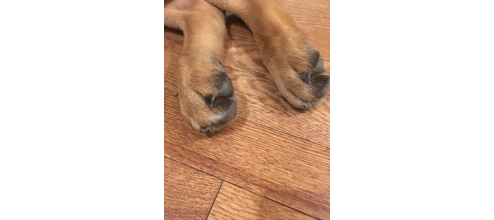
The back legs will start to cross over frequently and dogs will have a noticeably narrowed hind limb stance. Weakness is unfortunately progressive and many dogs may lose the ability to walk within 1 year after the symptoms develop. The condition is not considered painful but can lead to frustration for the pet as their mobility becomes more and more compromised. Sadly, there is no known cure for the disease.
Physical therapy for dogs with degenerative myelopathy
Dogs with degenerative myelopathy benefit from physical therapy. Studies have shown that dogs with physiotherapy remain ambulatory for longer than dogs without physical therapy.
A study of dogs assigned to no physiotherapy, moderate physiotherapy, and intensive physiotherapy showed that the dogs undergoing intense physiotherapy lived longer and remained ambulatory for longer ( 255 days versus 130 days for moderate physiotherapy and only 55 days for dogs with no physiotherapy).
Physiotherapy programs improve outcomes for dogs with degenerative myelopathy
The intense physiotherapy program for canine degenerative myelopathy consisted of:
- Slow walking with a sling as needed for 5 to 20 minutes 3-5 times per day.
- Dogs were encouraged to sit and get up several times a day. During walks, attention is placed on correct placement of paws. Full weight bearing on either hind limb was encouraged by pushing gently on the hip on each side.
- Passive range of motion exercises: gentle slow extension and flexion of each joint of both hind limbs starting distally and manipulating each joint separately.
- Passive range of motion was performed 3 times daily
OR
- daily hydrotherapy by walking on an underwater treadmill or swimming/ walking in water using assistance as needed for 5- 20 minutes
Moderate physiotherapy consisted of:
- Slow walking for 5-20 minutes three times per day
- Hydrotherapy or massage once a week. Massage was started as gentle stroking followed by kneading of the entire paravertebral muscles and limbs from distal to proximal.
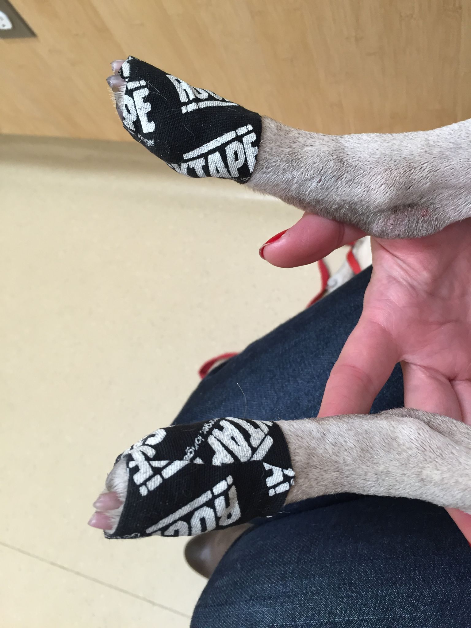
Protecting your dog’s feet from excess scuffing
As dogs with degenerative myelopathy are often scuffing their feet and wearing down their nails, this can cause ulcerations and bleeding. It is recommended to protect the paws by wrapping the feet in kinesiology tape or use booties.
Degenerative myelopathy can be prevented by testing for the mutation in breeding pairs of dogs. No breeding should occur with a dog who has a copy of the mutation.
Causes of acute back leg weakness in dogs
Acute weakness in the back legs is most commonly due to intervertebral disc disease (type I), spinal stroke, toxin exposure, immune mediated neurological disease, or severe systemic illness.
Acute IVDD (Type I) in dogs
Unlike Type II IVDD which results in more gradual degeneration, Type I IVDD develops rapidly. It occurs more commonly in small breed dogs like Dachshunds, Pekingese, Shih Tzus Toy/mIniature Poodles, Cocker Spaniels, Basset Hounds, chihuahuas and beagles. These dogs are typically of a young age (age 3 or older).
Type I disc disease occurs within minutes secondary to an acute rupture of the spongy disc that cushions the vertebrae. It is characterized by a sudden loss of normal movement function of the legs and pain.
Read this article for more information about Type II IVDD in dogs.
FCE (fibrocartilaginous embolism) in dogs
A type of “stroke” to the back legs that can also happen acutely is called a fibrocartilaginous embolism (FCE). It is caused by a small piece of the disc (the gelatinous center of the disc) which suddenly breaks off and travels in a spinal artery to an area in the back. This happens most commonly during a bout of vigorous exercise in a dog such as frisbee or ball play. FCE causes a rapid weakness that can progress to paralysis. The signs are typically lateralizing, meaning they are more likely to be apparent on one side more than another side.
This type of event happens most commonly in young to middle aged large breed dogs, but medium and small breed dogs can also be affected. Unfortunately there is no specific therapy other than supportive care. Dogs typically start to make improvement in their weakness and leg function within 1-2 weeks although full recovery can take many weeks longer. Some dogs have lasting weakness or neurologic deficits. Prognosis is based on the state of neurologic dysfunction at the outset of the event. Dogs who lose deep pain sensation have a very guarded prognosis for return to normal function, however, recovery is reported to be up to 80% in dogs.
Distinguishing IVDD from FCE in dogs
Acute IVDD and spinal strokes have obvious weaknesses on exams with neurologic deficits present. A spinal stroke, or fibrocartilaginous embolism (FCE), is painful initially but typically subsides quickly. Acute IVDD presents with more consistent pain. Both will cause similar neurologic deficits that lateralize mostly to one side (although both limbs can be affected). FCE classically occurs in large breed dogs or miniature Schnauzers while type I IVDD occurs more commonly in small breed dogs.
Systemic illness causing weakness in dogs and collapsing back legs
Systemic diseases can also cause weakness in dogs that may manifest itself with difficulty rising or exercise intolerance. Endocrine diseases like diabetes mellitus can cause damage to the peripheral nervous system that supplies the back legs and this can lead to weakness and proprioceptive deficits. Cushing’s disease is an over activity of the adrenal glands and produces a natural steroid. This can cause weakness or lethargy, and other classic symptoms like increased thirst, urination, hunge, panting and a large pot bellied appearance with thin skin and poor coat quality.
Hypothyroidism is another endocrine disorder that can cause lethargy , exercise intolerance and occasionally peripheral neuropathy and weakness. Finally, any dog that is suddenly ill can become compromised and weak.
Rare causes of acute hind end weakness in dogs: toxicities, immune mediated disease of the nervous system
Toxin exposure to botulinum toxin causes an acute weakness that starts in the hind limbs and gradually progresses up the body. The weakness as it progresses will threaten a dog’s ability to swallow and even breathe. The toxin is often secondary to eating contaminated, raw meat containing the toxin Clostridium Botulinum.
Immune mediated diseases of the nervous system, like