
Many cats awake from anesthesia with dilated pupils. This is called mydriasis. The most common cause for dilated pupils in a cat following anesthesia is the use of an opioid as part of the anesthetic protocol. The opioid effects on the pupil should wane within 24 (occasionally up to 48) hours. If your cat’s eyes remain dilated for longer than 48 hours after anesthesia, you should have your veterinarian evaluate your cat. The two other top causes of sudden dilated pupils are: cortical blindness and hypertension. Read further to learn more about these conditions.

Table of Contents
Opioid use can cause cats eyes to stay dilated after surgery
Cats are unique in their response to administration of opioids. Most other species develop small pupils from opioid administration. Opioids block the parasympathetic innervation of the pupil in cats. Catecholamine release caused by morphine’s effect on the brain stimulates their pupils to dilate. Opiods like morphine or Dilaudid are commonly used as a pre-medication for anesthesia.
The pupil is the muscle of the iris and it constricts or dilates in response to both light and the autonomic nervous system. The size of your cat’s pupils depends not only on the amount of light the eye receives but also whether the pupil is being controlled by the parasympathetic or sympathetic nervous system.
The parasympathetic nervous system controls bodily functions under normal circumstances. When an animal is suddenly threatened or startled, the sympathetic nervous system takes over. Evidence of the sympathetic nervous system at play is sudden pupil dilation when an animal is startled.
To help relax or calm cats, Feliway ,is an odorless calming pheromone that can be plugged in as a diffuser.

Increased sympathetic tone to the eye causes pupils to dilate
Under normal conditions, dim light will cause the pupils to dilate to allow more light to enter the eye. When an animal is scared, their sympathetic “fight or flight” responses will kick into gear and this will cause the pupil to dilate. The parasympathetic nervous system is in control during rest and relaxation. During this time the pupils are normal to small in size.
The analgesic effects of an opioid will wane far before the pupillary effects wane in cats. A typical opioid given as a premedication for anesthesia provides pain relief for 6-8 hours. However, the opioid’s disruption of the parasympathetic innervation to the eye lasts longer. This causes cats to have dilated eyes for many hours after anesthesia.
If your cat’s eyes remain dilated for longer than 24-48 hours after anesthesia, you should evaluate your cat for vision changes. Although there will be some diminished visual acuity when the pupils are both dilated, your cat should still be able to function normally.
Signs of vision loss in a cat after surgery
If your cat is struggling to see you will notice their movements will change . A cat that is visually impaired may navigate the room by hugging the walls to allow their tactile sense to help guide their movement. They may walk with a more crouched posture and be reluctant to jump up and down from furniture etc.
Home tests to check for vision loss in your cat
If you suspect vision loss in your cat, a couple of easy tests of vision you can do at home are: the menace test and the cotton ball test.
The menace response test
The menace response is an involuntary blink/flinch in response to the threat of something contacting their eye . This test is done with your hand held up, palm facing your cat’s one eye, slightly off to the side. Use a quick wave-like gesture towards the one eye. This should look as if the palm will contact their eye, and a visual cat should automatically blink. When you perform this movement you do not want to create wind that can be detected. You should perform the menace test on each eye.
The cotton ball test
The cotton ball test involves taking cotton wool and dropping it from a height above your cat. The cotton wool should not make a sound when falling. Your cat will want to track the cotton if they can see the cotton ball falling.
The PLR and dazzle response tests
Finally, two other eye tests involve using a bright light to shine in your cat’s eyes. These are the pupillary light response test (PLR) and dazzle response test.
To assess a pupillary light response (PLR): shine a bright light into your cat’s eye (but make sure the light is far enough away that it won’t cause your cat to close it’s eye). Light shining into the eye should evoke constriction of that pupil and cause the other pupil to constrict as well .
Cortical blindness in cats following anesthesia or surgery
It is important to note that the PLR and dazzle reflexes are “subcortical reflexes”. This means that these reflexes only test the pathway from the retina, optic nerve and midbrain. If an animal is blind from a problem in the visual cortex (cortical blindness) the PLR and dazzle reflexes can still be present and normal!
Persistently dilated pupils and evidence of vision loss following anesthesia is most concerning for a syndrome called cortical blindness. Cortical blindness occurs most commonly following anesthesia or seizures and is thought to develop secondary to hypoxia to the brain. The good news is that most cases of post anesthetic blindness will have vision return within a few days.
Use of a spring-loaded mouth gag during surgery can increase the risk of cortical blindness in cats
A study of 20 cats who developed cortical blindness following anesthesia determined that 16 of the 20 cats had a spring loaded mouth gag used as part of the procedure (either a dental procedure or an endoscopy where mouth gags were necessary to keep the mouth open for the treatment). Seventy percent of cats had recovery of their vision within 2 weeks.
Mouth gags were cited as a potential risk factor for cerebral hypoxia. The hypothesis was that use of a mouth gag occluded the maxillary artery and diminished important blood flow to the brain.
Cats are unlike other species and their cerebral blood flow depends on a maxillary branch of the carotid artery that runs along their jaw. When their mouth is stretched open too far, the bony projection on the edge of their mandible may pinch the maxillary artery and occlude blood flow.
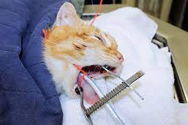
An example of a spring loaded mouth gag is shown below . The springs put constant pressure on the jaw to distract the mouth wide open.
A better solution is to use a shorter, non spring loaded mouth gag as shown in the other picture below.

Another cause for bilaterally enlarged pupils in a cat is high blood pressure
High blood pressure in cats can damage the retinas and this can lead to bilaterally enlarged pupils. Depending on the degree of damage, the cat may still have a pupillary light reflex and dazzle reflex. A fundic examination (retinal exam) may reveal multi focal retinal hemorrhages early in the disease. As time goes on, partial to complete retinal detachments can occur causing blindness.
Systemic hypertension in cats most commonly occurs secondary to kidney disease, hyperthyroidism or diabetes mellitus. A blood pressure measurement over 180mmHg combined with compatible ocular lesions is considered diagnostic. Blood pressure can be controlled with antihypertensive medication and in many cases vision can return within 6-8 weeks if blood pressure is adequately controlled.
Conclusion
It’s not uncommon for cats to come home from anesthesia with dilated pupils. This can last for many hours and sometimes may take 1-2 days to fully wear off. However, if you notice vision changes and dilated pupils, you should have your cat examined by your veterinarian.
There is an increased risk for sudden blindness in cats that had a spring loaded mouth gag utilized during an anesthetized procedure like a dental or endoscopy. Luckily, most cats will have their vision return to normal within 2 weeks.
The other most common cause for sudden pupil dilation and vision change in a cat is from high blood pressure damaging the eyes. This has nothing to do with anesthesia, but can still produce similar symptoms. A blood pressure measurement and evaluation of your cat’s retinas as a simple exam room test can easily diagnose this condition.
Depending on how severe the retinal changes are, some cats can have their vision return following several weeks on appropriate blood pressure medication.