
If your dog’s eyes appear to be bulging there are a number of possible causes. The eyes can be bulging due to problems with the size and positioning of the globe, or the tissues around the eye (such as the eyelids or muscles) can make the eyes appear to be bulging or swollen.
Read on to learn how to determine what is causing your dog’s eyes to bulge and what can be done to address this.

Optixcare lubricating eye drops are recommended by veteirnary ophthalmologists to help lubricate and keep red, irritated eyes comfortable.
Table of Contents
Some dogs’ eyes bulge naturally
Conformational bulging eyes. We all know that the cute brachycephalic breeds of dogs have bulging eyes naturally. This is because their boney orbits are more shallow compared to dolichocephalic breeds (longer nosed dogs. ) If the eye socket is more shallow, more of the globe will be pushed forward. The neonatal appearance of brachycephalic dogs (wide forehead, enlarged eyes and big head) provide some of the appeal of why breeds like Frenchies and Pugs are so popular. Unfortunately they can lead to a host of ophthalmologic problems in these breeds.
Brachycephalic ocular syndrome
Brachycephalic ocular syndrome is the collection of eye problems that brachycephalic breeds are prone to experiencing. Pugs, for example, have eye related medical disorders as the number one most common medical issue they face. In addition to increased rates of proptosis, these breeds commonly suffer from corneal trauma due to the fact that there is less bony protection and due to their decreased sensation of their cornea. They sometimes have a problem called trichiasis (eyelashes rubbing on the cornea) ,often because their lower eyelids can roll inwards. These dogs frequently develop pigmentation on their corneas from chronic exposure irritation.
The orbit where the globe of the eye sits is completely encased in bone and cannot expand. When there is a problem with the globe of the eye, it may be pushed forward (exophthalmia), out of the socket (proptosis), or distorted and enlarged (buphthalmia).
Bulging eye problems in dogs related to a problem with the globe
| Diagnosis | Globe | Orbit |
| Proptosis | Normal | Normal |
| Exophthalmia | Normal | Abnormal |
| Buphthalmia | Abnormal | Normal |
Traumatic eye bulging in a dog
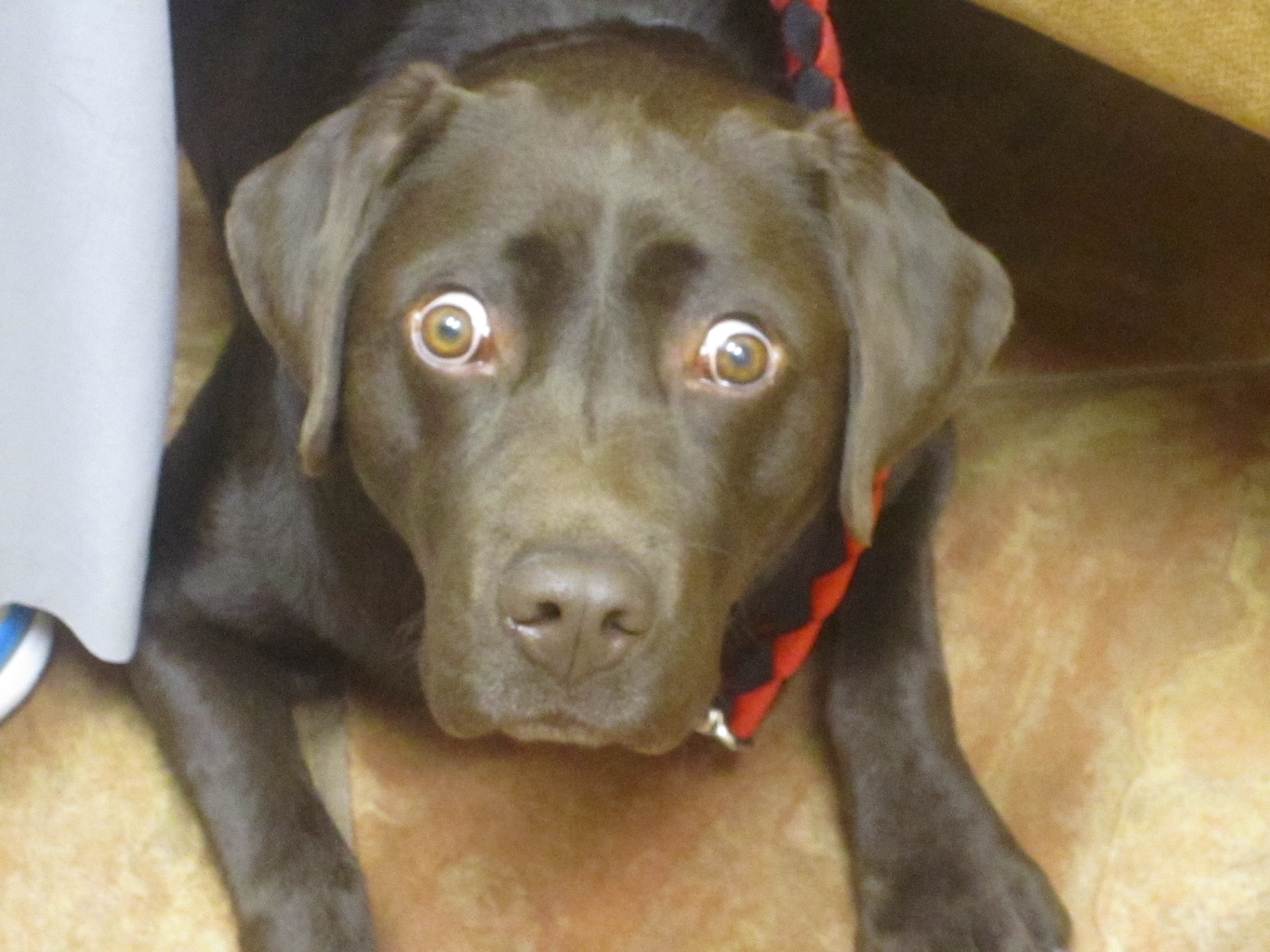
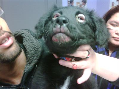
The most obvious cause of a bulging eye (and perhaps the most scary) is called proptosis. This is caused by trauma which pushes the globe forward in front of the eyelids. With a proptosed eye the eyelids will not be visible all the way around the eye.

Today’s Veterinary Practice discusses here all about proptosis. This photo shows a proptosed eye in a dog. The globe is pushed forward and the eyelids are trapped behind the “equator” of the glove.
Brachycephalic breeds are most at risk for proptosis
Brachycephalic breeds of dogs are most at risk for proptosis because their boney orbits are much more shallow. Even a more minor scuffle can increase pressure enough to pop out an eyeball. While this is scary, they are also more likely to have less surrounding muscle trauma or nerve damage because less force is necessary to proptose the eye. These dogs are far more likely to retain vision after replacement of the globe within the orbit.
Proptosis in dogs in a medical emergency
Proptosis is a medical emergency. In many dogs, the trauma to the eye can tear the supporting muscles that attach to the globe of the eye. After the eye is replaced into the globe, there may be a permanent strabismus (the eye may always be looking aside) because the supporting muscles that attach to one side may be torn. There is also the risk of vision loss if the optic nerve (the large nerve which is attached to the back of the eye is stretched from the trauma. Finally, the clear surface of the eye (cornea) may be damaged requiring special treatment.
Exophthalmia in dogs
Exophthalmia (globe being pushed forward) can occur from a space occupying process (mass, fluid etc. ) that has accumulated behind the eye. This will put pressure on the globe and make the eye bulge.
An examination tip to determine if your dog is suffering from exophthalmia
A quick examination tip can help determine if this is a possibility. This is called a retropulsion test. Retropulsion is soft but gentle pressure on the closed eye. They should be able to gently push the eye back into the socket. If there is a space occupying mass or fluid behind the eye, retropulsion will not be possible or more difficult.
Retrobulbar abscesses in dogs (abscesses behind the eye)
Retrobulbar abscesses can occur in dogs most commonly secondary to a deep tooth root abscess or due to a penetrating foreign body like a stick or plant material and migrates through the back of the mouth into the palate and creates an infection in the back of the eye.
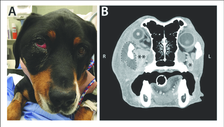
The dog featured in this publication had a retrobulbar abscess due to migrating plant material that entered behind a tooth and created an infection behind his eye.
Clinical signs of this will be one-sided exophthalmos with swollen conjunctiva, a red inflamed eye, ocular discharge, and an elevated third eyelid may be present. There is often pain on opening the mouth wide and a swelling is sometimes notable in the back of the mouth near the roof of the mouth.
How to diagnose an infection behind the eye in a dog
The diagnosis is based on imaging (a CT scan of the head is best) and then the area is drained and flushed under general anesthesia. A culture will determine what bacteria are infecting the abscess. Nocardia or actinomyces are bacterial infections that may need weeks of antibiotic therapy and are best eliminated if the wound/abscess is surgically debrided. If an infection in this area is drained but recurs, then an exploratory surgery to open up the site is recommended to look for and remove possible trapped foregin material.
Bulging eyes due to extraocular muscle inflammation
Bulging eyes due to extraocular myositis (EOM) in dogs
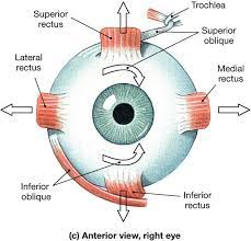
Eyes may bulge if there is a disease causing marked inflammation and swelling of the muscles surrounding the eye. This is called extraocular myositis (EOM). EOM is an immune mediated disease of the extraocular muscles. The exact mechanism is unknown but the condition is a targeted attack on specific antigens present only on the muscles that control eye movement (rectus and oblique extraocular muscles). The specific targeting of only these muscle groups is thought to somehow be related to their embryologic origin.
Exam findings for extraocular myositis in dogs
EOM causes a non-painful bilateral exophthalmos. The whites of the eyes are very visible and there is no third eyelid elevation. There may be retraction of the upper eyelids and some congestion of the vessels on the sclera (whites of the eye). Both globes should retropulse normally but return to their original position afterwards. There should be no pain on opening of the mouth. There is occasionally some vision loss if there is enough swelling of the optic nerve in the back of the eye.
Canine characteristics for extraocular myositis
- Large breed dogs are over-represented
- More commonly affects dogs under the age of one
- Females are twice as likely to develop this disease
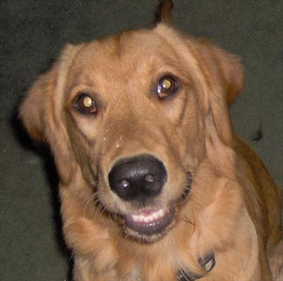
Predisposed breeds affected by extraocular myositis
Golden retrievers
Labrador retriever
Akitas
Dalmations
Chinese shar pei
Irish Wolfhound
English springer spaniel
Doberman pinschers
Diagnosing and treating extraocular myositis in dogs
Technically a formal diagnosis is made with biopsy of the muscle tissue to diagnose the specific type of inflammation in the tissue. Most of the time the diagnosis is made on physical exam findings alone because the symptoms are very specific and biopsying the muscles affected is invasive.
Most dogs respond to immunosuppressive doses of steroids : prednisone 1-2mg/kg q 12 hours and tapered after 3 weeks. Some dogs require long term treatment or repeated treatments.
Symptoms begin to improve quickly within 1 week in most cases. Many cases studied showed no recurrences after 10 months of monitoring. Chronic extraocular myositis that is not treated can lead to scarring of the muscle tissue and chronically deviated eyes (strabismus).
Bulging eyes due to masticatory muscle myositis in dogs
Masticatory muscle myositis is a condition that causes inflammation of the muscles that control chewing (mastication). IT is the most common cause of inflammatory muscle disease. Type 2m muscle fibers are primarily in the muscles of mastication. Autoantibodies target Type 2m muscle fibers.
Exam findings in dogs with masticatory muscle myositis (MMM)
The underlying triggers for this immune dysregulation is not known. Physical exam findings include a reluctance to open the mouth and may be the first sign noted.
My first case of this disease in practice was a young boxer that presented to me because her astute owner noticed that she stopped wanting to carry around her favorite balls. On her exam, when I tested opening her mouth, she yelped in discomfort. Following treatment she was back to carrying around her tennis balls.
Examination may show swelling along the master and temproalismiscles on the cheek areas. Dogs with chronic disease may have muscle atrophy that is typically bilateral. A diagnosis can easily be made by submitting a blood panel for type 2m autoantibodies.
Characteristics of dogs with masticatory muscle myositis
Any age, breed or sex of dog can be affected. Larger breeds of dogs are more commonly affected. The golden retriever, labrador retriever, German shepherd dog, rottweiler, weimaraner and doberman pinscher are over-re-reatend. Most dogs are mature adults, but the average age is 3-4 years.
Eye signs are seen in upto 44% of dogs with this disease. These eye signs develop secondary to the swelling or later atrophy of the pterygoid and temporalis muscles. This can cause exophthalmos and pain on globe retropulsion, third eyelid elevation and eye discharge.
Treatment of dogs with masticatory muscle myositis
In the acute stage MMM response to immunosuppressive doses of steroids again at 1-2mg/kg orally every 12 hours. This dose is maintained until there is normal jaw function (about 4 weeks) and then slowly tapered every month by 50%. Steroids are usually given for 6-8 months to decrease the risk of relapse.
Eyelid swelling in dogs: blepharitis
Blepharitis is inflammation of the eyelids that causes swelling and often a secondary bacterial infection. It can be bilateral or unilateral. The most common underlying cause is allergic skin disease followed by infectious/parasitic disease. Less common causes of blepharitis are due to immune skin diseases such as pemphigus, discoid lupus or uveodermatologic syndromes in dogs.

Allergic bacterial eyelid infection in dogs
Allergic blepharitis (atopic dermatitis) may cause swelling and secondary bacterial infection of the eyelids. Your veterinarian can perform a cytology of the affected skin and determine if there is a bacterial infection present. The presentation is typically bilateral eyelid swelling and ulceration. The treatment is typically a combination of an oral antibiotic and a topical or oral steroid to reduce the inflammation and swelling. Eyelids can be gently cleaned with an eyelid wipe like Systane lid wipes available on Amazon.
Eyelid swelling due to demodex infection in dogs
Parasitic blepharitis is most often secondary to demodex mite infection and typically presents in puppies younger than 10 months of age. This can present only on one eye and there is often hair loss around the eye, mild redness and scaling. Demodex can be treated with oral flea/tick preventatives Nexgard and Bravecto. If your dog is already on one of these products, their blepharitis should not be due to a demodex infection.
Puppy strangles: juvenile cellulitis
Juvenile sterile cellulitis is an immune mediated skin disease that is of unknown etiology . It occurs in young dogs, primarily puppies. It causes a swollen face, particularly around the eyelids, lips and muzzle with enlarged submandibular lymph nodes. Within 24-48 hours, little red bumps and pustules can appear on the lips, muzzle, chin , nose and around the eyes. Half of affected puppies will be lethargic with a fever and decreased appetite. This condition responds to immunosuppressive doses of steroids at 1mg/kg twice a day for 2-4 weeks. Systemic antibiotics may also be needed if there is a secondary bacterial infection.
Buphthalmia is a stretched out globe
The most common cause of a buphthalmic globe is increased pressure within the eye called glaucoma.
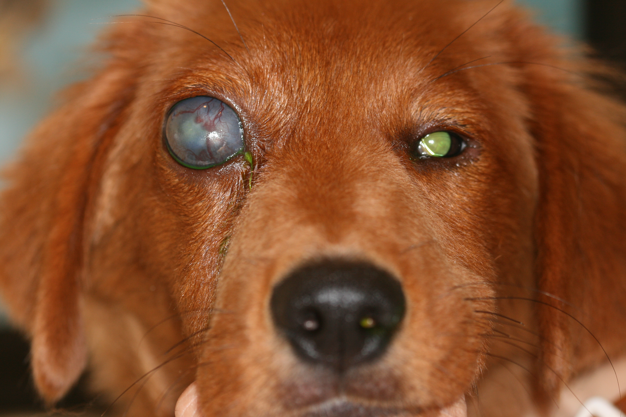
You can distinguish buphthalmia from exophthalmos because the diameter of the cornea is not the same in both eyes. In contrast to exophthalmos, where the globe is pushed forward, the conjunctiva in a buphthalmic patient are not inflamed or swollen. Dogs with exophthalmos often have an irritated, swollen conjunctiva and a normal intraocular pressure.
Buphthalmia is caused by chronic glaucoma causing long standing elevated pressures inside the eye.This is from an imbalance between the production of aqueous fluid in the eye and the normal drainage of this fluid. Glaucoma is identified by an increased pressure measured within the eye measured easily by instrumentation your veterinarian will have at the clinic. By the time the globe has stretched this far, no treatment is possible to restore the eye to a normal appearance. Some cases of glaucoma are unfortunately intractable to glaucoma therapies.
Bulging eyes in dogs conclusion
If your dog has bulging eyes, the problem can be related to the eyeball itself (the location of the globe), the size of the globe, or the periocular tissues of the eye which may be swollen.